| Cat # | Size | Price | Quantity | |
|---|---|---|---|---|
| 204003 | 25 tests | $80 | ||
| 204004 | 100 tests | $180 |
| Clone | M5/114.15.2 |
|---|---|
| Application | Flow Cytometry |
| Reactivity | Mouse |
| Format | iF647 |
| Target Name | I-A/I-E, MHC class II |
| Isotype | Rat IgG2b |
| Antibody Type | Monoclonal |
| Regulatory Status | RUO |
| Formulation | Phosphate-buffered solution, pH 7.2, containing 0.09% sodium azide and 0.2% (w/v) BSA |
| Protein Concentration | Supplied at a lot-specific concentration. |
| Storage&Handling | The antibody solution should be stored undiluted between 2°C and 8°C, and protected from prolonged exposure to light. Do not freeze. |
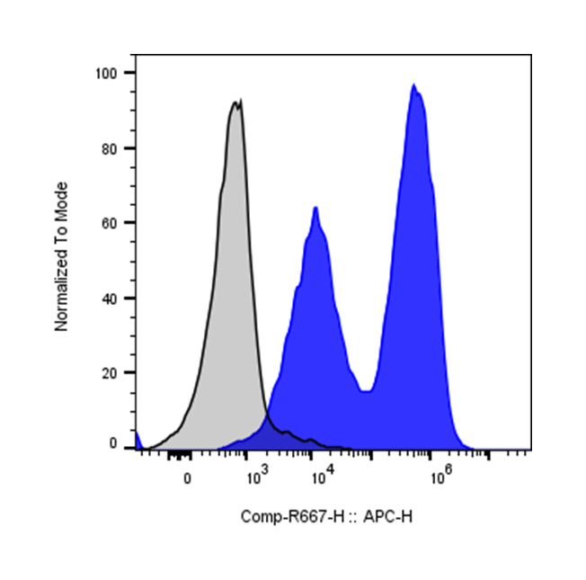
| Recommended Usage | For flow cytometric staining, it is recommended to use 5 µL of this reagent per 0.5-1.0 million cells in a 100 µL volume. Optimal reagent performance should be determined by titration for each specific application. iF647 has an excitation max at 656 nm and an emission max at 670 nm. |
| Excitation Laser | Red Laser (633 nm) |
| See All Formats | Clone M5/114.15.2 |
Mouse I-A and I-E are the classical major histocompatibility complex (MHC) class II molecules in mice. They are encoded within the murine MHC locus, also known as the H-2 complex, and function as key antigen-presenting molecules in the adaptive immune system. These proteins are primarily expressed on professional antigen-presenting cells such as dendritic cells, macrophages, and B cells, although they can also be induced on other cell types during inflammation. Their primary role is to present processed extracellular peptide antigens to CD4+ T helper cells, thereby initiating and regulating immune responses against pathogens and other foreign antigens.
Structurally, I-A and I-E molecules are heterodimeric glycoproteins composed of an α chain and a β chain. Each chain contains two extracellular domains, a transmembrane region, and a short cytoplasmic tail. The peptide-binding groove is formed by the α1 and β1 domains and is open at both ends, allowing it to accommodate peptides typically 12–20 amino acids in length. This open structure distinguishes MHC class II molecules from class I molecules and permits binding of longer peptides with flexible extensions beyond the groove. Genetic polymorphisms in both the α and β chains influence peptide binding specificity and contribute to differences in immune responses between mouse strains.
The ligands presented by I-A and I-E are primarily peptides derived from extracellular proteins that have been internalized by antigen-presenting cells through endocytosis or phagocytosis. These proteins are processed within endosomal and lysosomal compartments into peptide fragments, which are then loaded onto MHC class II molecules with the help of accessory molecules such as the invariant chain and H-2M. The resulting peptide–MHC complexes are transported to the cell surface, where they are recognized by the T cell receptor (TCR) on CD4+ T cells, leading to T cell activation and cytokine production.
Mouse I-A and I-E molecules play important roles in susceptibility to autoimmune and inflammatory diseases in experimental models. Certain MHC class II haplotypes are associated with conditions such as experimental autoimmune encephalomyelitis (a model for multiple sclerosis), collagen-induced arthritis, and type 1 diabetes in non-obese diabetic (NOD) mice. Differences in peptide presentation can influence the activation of autoreactive T cells and thus determine disease susceptibility or resistance.
Because of their central role in antigen presentation, I-A and I-E molecules are important in immunological research and therapeutic development. They are widely used in mouse models to study T cell responses, vaccine mechanisms, and autoimmune pathogenesis. In therapeutic contexts, strategies that alter peptide presentation or modulate CD4+ T cell activation—such as peptide-based tolerizing vaccines or antigen-specific immunotherapies—often rely on understanding how peptides interact with MHC class II molecules like I-A and I-E. As a result, these molecules remain fundamental tools for studying immune regulation and designing new immunotherapies.
iF647 Rat IgG2b Isotype Control Antibody
iF647 Anti-mouse I-A/I-E Antibody TDS
Have a product or application question? Consult our FAQs or contact us.