| Cat # | Size | Price | Quantity | |
|---|---|---|---|---|
| 112103 | 25 tests | $75 | ||
| 112104 | 100 tests | $210 |
| Clone | GL7-mM |
|---|---|
| Application | Flow Cytometry |
| Reactivity | Human, mouse |
| Format | iF647 |
| Target Name | T and B cell activation markerT and B cell activation marker, Ly77 |
| Isotype | mouse IgM |
| Antibody Type | Monoclonal |
| Regulatory Status | RUO |
| Formulation | Phosphate-buffered solution, pH 7.2, containing 0.09% sodium azide and 0.2% (w/v) BSA |
| Protein Concentration | Supplied at a lot-specific concentration. |
| Storage&Handling | The antibody solution should be stored undiluted between 2°C and 8°C, and protected from prolonged exposure to light. Do not freeze. |
| Recommended Usage | For flow cytometric staining, it is recommended to use 5 µL of this reagent per 0.5-1.0 million cells in a 100 µL volume. Optimal reagent performance should be determined by titration for each specific application. iF647 has an excitation max at 656 nm and an emission max at 670 nm. |
| Excitation Laser | Red Laser (633 nm) |
| See All Formats | Clone GL7-mM |
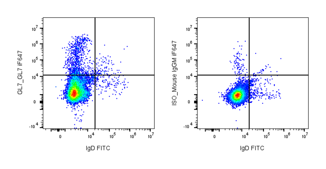
GL7 antigen is a cell-surface carbohydrate epitope commonly used as a marker of activated B cells, particularly germinal center (GC) B cells. It was originally identified through the monoclonal antibody GL7, which recognizes a specific glycan structure expressed on lymphocytes during immune activation. In both mouse and human immunology research, GL7 expression is widely used to identify germinal center B cells in secondary lymphoid organs such as lymph nodes, spleen, and Peyer’s patches. These cells are actively undergoing proliferation, somatic hypermutation, and affinity maturation during adaptive immune responses.
Structurally, GL7 is not a classical protein antigen but rather a glycan epitope present on glycoproteins and glycolipids at the cell surface. The epitope corresponds to a modified sialylated lactosamine carbohydrate structure that becomes exposed when certain sialic acid residues are enzymatically altered during B-cell activation. Because the GL7 determinant is carbohydrate-based, it may appear on multiple membrane proteins or lipids that carry the appropriate glycosylation pattern. This explains why GL7 is considered a differentiation marker rather than a receptor with a single defined protein backbone.
Unlike classical immune receptors, GL7 does not have a well-defined ligand or signaling function. Instead, it reflects changes in cellular glycosylation associated with activation and germinal center differentiation. The appearance of GL7 correlates with B-cell interactions with T follicular helper cells and antigen stimulation within germinal centers. As a result, GL7 is often used together with markers such as B220, CD95 (Fas), and IgD to identify germinal center B-cell populations by flow cytometry or immunohistochemistry.
GL7 expression is associated with immune responses and can also appear in pathological conditions involving abnormal germinal center activity. For example, elevated germinal center reactions marked by GL7-positive B cells may occur in autoimmune diseases, chronic infections, or certain B-cell lymphomas. Although GL7 itself is not typically targeted therapeutically, it is widely used in research and preclinical studies to monitor germinal center responses, vaccine-induced B-cell activation, and immune dysregulation. In this way, GL7 serves as an important biomarker for studying adaptive immune responses and evaluating immunotherapies or vaccine efficacy.
iF647 Mouse IgM Isotype Control Antibody
iF647 anti-mouse/human GL7 Antigen Antibody TDS
Anti-Mouse/human CD45R/B220 Antibody, Clone RA3-6B2
PE/Cyanine7 Anti-Mouse/human CD45R/B220 Antibody, Clone RA3-6B2
iF647 Anti-Human CD95 (Fas) Antibody, Clone DX2
Have a product or application question? Consult our FAQs or contact us.